Suite 5, Level 1
66 Pacific Hwy
ST LEONARDS NSW 2065
66 Pacific Hwy
ST LEONARDS NSW 2065
Fax: 02-8412 0060


Diabetes is a condition that affects the ability of the body to use and store sugar. There are two types of diabetes:
Symptoms from diabetes are caused by high levels of blood sugar and may include:
These symptoms may occur suddenly, may develop over months or years, or there may not be any symptoms.
As the body does not use and store sugar correctly, it can cause high blood sugar levels. This creates changes in the body’s veins, arteries and capillaries that carry blood throughout the body.
After 10 or 15 years of diabetes, most people develop signs of mild damage to the retina at the back of the eye, known as diabetic retinopathy. However this may have no effect on vision. Retinopathy can affect sighti f it becomes significant or severe. Therefore it is a good idea to have regular eye checks at least every 1-2 years.
The 4 main eye problems associated with diabetes are focusing problems, cataracts, glaucoma and diabetic retinopathy.
This is common in people who have just been diagnosed with diabetes and have commenced treatment. Diabetes can cause fluctuating vision in both eyes. As the blood sugar level rises and falls, the tissues of the eye can cause fluid to pass intothe lens, making it swell and become thicker. When the lens becomes thicker, it only allows the eye to focus on objects at near, and images will be out of focus.Good control of blood sugar levels will minimise episodes of blurred vision. The problem may occur on and off for some time but usually settles in about one month after starting diabetic treatment.
The lens inside the eye is normally clear, however if a cataract forms, the lens becomes cloudy and can affect vision. Cataracts also scatter ilght, leading to a sensation of glare. Cataracts are common in older people, but can also develop earlier in those with diabetes. Eventually cataract surgery may be required to clear the vision by removing the cloudy natural lens inside the eye and replacni g it with a clear artificial lens.
If blood vessels in the eye become blocked, the eye produces tiny new blood vessels (neovascularisation). The new blood vessels grow along the surface of the retina, however the neovascularisation can also occurin the front of the eye on the iris. This can block the drainage mechanism of the eye so the fluid cannot flow out of the eye. This causes a build-up of pressure (neovascular glaucoma), which can cause damage to the optic nerve and can result in blindness.
Diabetic retinopathy (DR) occurs when the very fine blood vessels in the retina are damaged due to diabetes. The longer a person has diabetes, the greater the risk of DR.
There are 2 main types of diabetic retinopathy: non-proliferative diabetic retinopathy and proliferative diabetic retinopathy, depending on whether or not new blood vessels (neovascularisation) have started to grow. These new blood vessels indicate very severe disease.
This is the early stage of DR, which involves tiny areas of bleeding of blood vessels or leaking of fluid or fats into the retina. The fluid can cause the retina to swell m( acular oedema) and the fats that have escaped from the blood vessels may form deopsits (exudates) on the retina. At this stage treatment is usually not necessary as long as the macular (central part of the retina) is not involved. This early stage should be closely monitored and blood sugar levels should be controlled.
Signs of NPDR may include:
Retinal Haemorrhages: tiny spots of blood that leak into the retina but are rarely responsible for any loss of vision by themselves.
Microaneurysm: small out pouching in the retinal blood vessels that often leak fluid.
Hard Exudates: deposits of cholesterol or other fats from the blood that have leaked into the retina.
At this stage visual problems are usually not noticed and vision may not change until the disease is advanced. Treatment is usually not required as long as the macula (thecentral part of the retina) is not involved. Regular eye exams (every 1-2 years) will reduce the risk of vision loss and blindness and can delay the development of retinopathy. This early stage should be closely monitored and blood sugar levels should be ocntrolled.
As diabetic retinopathy worsens, blood vessels in the retina become blocked due to diabetes and new blood vessels may grow or ‘proliferate’ on the surface of the retina or optic nerve, to replace them. These new vessels are fragile and can bleed easily into the vitreous jelly inside the eye (vitreous haemorrhage). Bleeding can cause scar tissue to form between the vitreous and the retina, and can pull the retina out of position acusing it to become detached. This is calledtractional retinal detachment and may need urgent treatment. When retinal detachment occurs, people may notice a sudden loss of vision or a dark shadow.
The macula is the central part of the retina, which is responsible for clear vision. The most common type of retinopathy affecting vision is due to swelling of the macula, which is caused by leaking fluids from the retinal blood vessels. If left untreated the centre of vision is permanently damaged. This will affect your ability to read. Laser treatment is recommended for the treatment of macular oedema.
The fragile new blood vessels may bleed into the vitreous. This may prevent the light rays from reaching the retina. If the haemorrhage is small, a person may only see a few dark spots but if the haemorrhage is large, this may significantly obstruct vision, allowing only light and dark perception.
The haemorrhage may take days, months or even years to be reabsorbed into the bloodstream, depending on the amount of blood present. When the blood clears, vision may return to normal unless the macula is damaged. However, if the eye on its own cannot adequately reabsorb the blood within a reasonable time, then vitrectomy surgery may be required. In this surgery the ophthalmologist removes the vitreous jelly containing blood from the inside of the eye, and replaces it with a clear saline solution.
When PDR is present, the scar tissue associated with neovascularisation may shrink, wrinkle or pull the retina from its normal position.
If macular wrinkling occurs, this causes vision distortion. Vision can be severely affected when the macula or larger areas of the retina are detached. The longer the maculais distorted or left out of place the more severe the vision loss will be. Vitreous haemorrhage may be recurrent and blood may accumulate faster than the eye can reabsorb it.
People who are at most risk are those:
It is important to realise that anyone who has ever been diagnosed with diabetes is at risk, even if diabetes is controlled with diet alone.
When to schedule an eye examination
People with diabetes should see their ophthalmologist immediately if they have noticed any visual changes. Otherwise, eye examinations should be scheduled at least once a year.
Juvenile onset or insulin dependent patients should see their eye doctor every yeafrrom 10 years of age and onwards.
Anyone over the age of 30 years who has just been diagnosed with diabetes should have an eye examination within a few months of diagnosis. Diabetic retinopathy may have already occurred if diabetes has been undetected fora while.
Pregnant women with diabetes should schedule an appointment with their ophthalmologist in their first trimester because retinopathy can progress quickly during pregnancy.
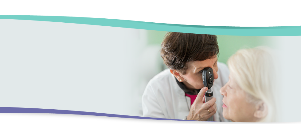
An ophthalmologist can often diagnose and treat seirous retinopathy before you are aware of any vision problems.
DR is detected during an eye examination of the retina through enlarged (dilated) pupils and by testing your vision. You will have drops in your eyes that dilate the pupils and the ophthalmologist will examine your retina with special instruments. When pupils are dilated, vision may be blurred for a few hours and glare will also be annoying from bright lights during that time.
If your retina appears to be affected, fluorescein angiography may eb recommended. This is a procedure used to further evaluate the retina. A yellow dye is injected into a vein in the arm. The dye enters the bloodstream and flows through the retinal blood vessels. A series of photographs are taken of the retina using a special camera. The dye indicates any leaking of blocked blood vessels and can indicate whether neovascularisation is beginning. It also gives an indication of whether laser treatment is needed and where it should be applied.
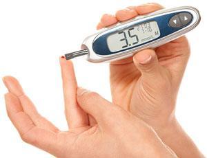
The best treatment is to prevent the development of retinopathy as much as possible. Strict control of blood sugar levels and blood pressure play an important role in reducing the long-term risk of vision loss from diabetic retinopathy.
Laser is often recommended for people with macular oedema, advanced retinopathy and neovascular glaucoma. It is used to treat retinopathy and is very effective in maintaining vision but cannot restore vision that has already been lost. Laser treatment ami s to seal leaking blood vessels in the retina, prevents further damage, and reduces the risk of vision loss.
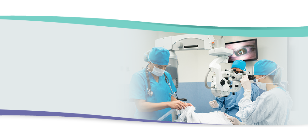
Vitrectomy surgery is indicated if there is blood in the vitreous that is affecting vision (and hasn’t cleared away), and if there is retinal scar tissue. During this operation, the vitreous contaminated with blood and retinal scar tissue that is present and causing a problem are removed and replaced with clear fluid. This fluid is gradually absorbed and replaced by fluid produced by the eye. In some cases a special gas or silicone oil may be applied inside the eye.